Digital X-Ray & Radiography Services for Pets in Fairfield, TX
The Power of Digital Radiography in Veterinary Medicine
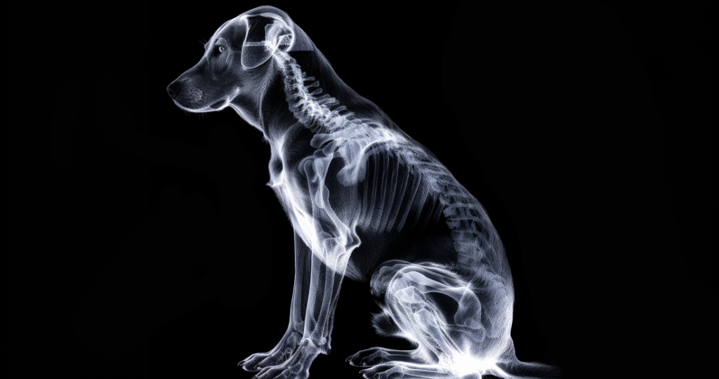
Digital radiography has revolutionized how veterinarians diagnose and treat internal conditions that remain invisible during physical examinations, making pet x-ray Fairfield TX services essential for comprehensive veterinary care. Unlike traditional film x-rays that required chemical processing and extended waiting times, digital radiography provides instant, high-resolution images that can be enhanced, magnified, and shared electronically. This advanced technology at our Fairfield clinic allows us to identify problems ranging from broken bones to heart disease within minutes of image capture.
The transition from conventional film to digital radiography represents one of the most significant advances in veterinary diagnostic imaging over the past decade. Digital sensors capture x-ray images with greater detail and clarity than film, revealing subtle abnormalities that might have been missed with older technology. The ability to adjust contrast, brightness, and magnification after image capture means we can optimize visualization of different tissues without retaking x-rays, reducing your pet’s radiation exposure.
Our investment in state-of-the-art digital radiography equipment demonstrates our commitment to providing Central Texas pets with the highest quality diagnostic services available. The immediate availability of images means faster diagnosis, quicker treatment initiation, and better outcomes for our patients. This technology proves invaluable for both emergency situations requiring rapid assessment and routine screening for early disease detection.
How Digital X-Rays Work and Their Advantages
Digital radiography uses electronic sensors instead of traditional photographic film to capture x-ray images, similar to how digital cameras replaced film cameras. When x-rays pass through your pet’s body, different tissues absorb varying amounts of radiation based on their density. Dense structures like bones appear white, soft tissues show as various shades of gray, and air-filled structures appear black on the resulting image.
The digital sensors in our pet x-ray Fairfield TX equipment are far more sensitive than traditional film, requiring less radiation to produce diagnostic quality images. This increased sensitivity means safer imaging for your pet, especially important for young animals or those requiring multiple x-rays. The reduced radiation exposure also provides added safety for our veterinary staff who work with imaging equipment daily.
Image processing capabilities of digital systems allow enhancement techniques impossible with traditional film. We can adjust window and level settings to optimize visualization of specific tissues, apply filters to enhance edge detection, and measure structures precisely using digital tools. These capabilities often reveal pathology that would have required additional views or even different imaging modalities with conventional radiography.
Common Conditions Diagnosed with X-Rays
Skeletal system evaluation remains one of the primary uses for radiography in veterinary medicine. Fractures, from simple breaks to complex multi-fragment injuries, are immediately visible on x-rays, allowing us to plan appropriate treatment. Arthritis and degenerative joint disease show characteristic changes including bone spurs, joint space narrowing, and increased bone density that help guide pain management strategies.
Thoracic radiographs provide invaluable information about heart and lung health. Heart enlargement, fluid accumulation in lungs, pneumonia, and masses become visible through chest x-rays. The size and shape of the heart, visibility of blood vessels, and lung field patterns help diagnose conditions ranging from congestive heart failure to metastatic cancer.
Abdominal radiographs reveal problems with organs, detect foreign objects, and identify life-threatening conditions like intestinal obstruction or gastric dilatation-volvulus (bloat). While soft tissue structures are less distinct than bones on x-rays, abnormal gas patterns, organ displacement, and mineral densities provide diagnostic clues. Bladder stones, enlarged organs, and fluid accumulation all appear on abdominal radiographs.
Emergency Applications of Digital Radiography
Trauma cases require rapid radiographic assessment to identify all injuries and prioritize treatment. Our digital system’s speed proves life-saving when pets arrive after vehicle accidents, falls, or animal attacks. Within minutes, we can evaluate for pneumothorax (collapsed lung), internal bleeding, fractures, and organ damage, allowing immediate surgical intervention when necessary.
Foreign body ingestion represents a common emergency where x-rays provide crucial information for treatment decisions. While not all foreign objects are visible on x-rays, many items including bones, metal objects, and certain plastics appear clearly. The location and size of foreign objects determine whether surgical removal is necessary or if medical management might allow passage.
Respiratory emergencies benefit from immediate chest radiographs that reveal underlying causes of breathing difficulty. Pulmonary edema, pleural effusion, pneumonia, and tracheal collapse each show distinct radiographic patterns requiring different treatments. Our ability to obtain and interpret these images quickly means appropriate therapy begins without delay, improving survival rates for critical respiratory patients.
Orthopedic Imaging and Surgical Planning
Comprehensive orthopedic evaluation requires multiple radiographic views to fully assess bone and joint problems. Our digital system allows rapid positioning changes and immediate image review, ensuring we capture all necessary angles without keeping pets under sedation longer than necessary. Standard orthopedic studies include at least two perpendicular views of affected areas, with additional specialized views for specific conditions.
Pre-surgical planning for orthopedic procedures relies heavily on detailed radiographic evaluation. Digital measurements allow precise implant sizing for fracture repairs, while image manipulation helps visualize optimal surgical approaches. We can overlay templates for various surgical implants directly onto digital images, ensuring appropriate equipment is available before surgery begins.
Post-operative radiographs monitor healing progress and verify proper implant positioning:
- Immediate post-surgical images confirm correct placement of pins, plates, or screws
- Follow-up radiographs assess bone healing and callus formation
- Sequential imaging tracks healing progression over weeks to months
- Implant complications like loosening or migration are detected early
- Healing delays or non-unions are identified before clinical problems develop
- Weight-bearing recommendations are adjusted based on radiographic healing
- Implant removal timing is determined by complete radiographic healing
Dental Radiography for Complete Oral Health
Dental radiography reveals problems hidden below the gum line where most dental disease occurs. Our pet x-ray Fairfield TX capabilities include specialized dental sensors that provide detailed images of individual teeth and surrounding bone. Since 70% of each tooth lies below the visible crown, dental x-rays are essential for complete oral health assessment.
Periodontal disease evaluation through dental radiographs shows bone loss around tooth roots that determines whether teeth can be saved or require extraction. Tooth root abscesses, often causing facial swelling or draining wounds, become clearly visible on dental x-rays. Fractured teeth may have root damage requiring extraction even when crowns appear intact.
Feline tooth resorption, a painful condition affecting many cats, often goes undetected without dental radiographs. These lesions destroy tooth structure from within, causing significant pain despite minimal visible changes. Early detection through radiography allows treatment before severe pain develops, improving quality of life for affected cats.
Cancer Detection and Staging
Radiography plays a crucial role in cancer diagnosis and staging, helping determine treatment options and prognosis. Primary bone tumors like osteosarcoma show characteristic patterns of bone destruction and new bone formation. Early detection of bone cancer through radiography may allow limb-sparing surgery rather than amputation in some cases.
Metastatic disease evaluation includes chest radiographs to check for lung metastases, the most common site for cancer spread. The presence or absence of visible metastases significantly affects treatment recommendations and prognosis. Our high-resolution digital images can detect smaller metastatic nodules than traditional film, providing more accurate staging information.
Abdominal masses discovered during physical examination require radiographic evaluation to determine organ of origin and assess for spread. While additional imaging like ultrasound often provides more detail for soft tissue masses, radiographs offer valuable initial information about size, location, and effect on surrounding structures.
Contrast Studies and Special Procedures
Contrast radiography uses special dyes to enhance visualization of structures that normally appear similar on standard x-rays. Barium studies outline the gastrointestinal tract, revealing obstructions, foreign bodies, or abnormal motility patterns. These studies require multiple images over time, where digital radiography’s immediate results prove invaluable for determining when adequate contrast has reached different intestinal segments.
Urinary contrast studies including cystograms and urethrograms help diagnose bladder ruptures, urethral obstructions, and ectopic ureters. Intravenous contrast studies evaluate kidney function and ureter patency. Digital technology allows real-time monitoring of contrast flow, capturing images at optimal moments for diagnostic information.
Myelography, injection of contrast around the spinal cord, helps diagnose intervertebral disc disease and spinal tumors when advanced imaging isn’t available. Digital enhancement of myelogram images often reveals subtle compression not visible on standard views. While MRI has largely replaced myelography, this technique remains valuable when advanced imaging isn’t accessible or affordable.
Pregnancy Detection and Monitoring
Radiographic pregnancy diagnosis becomes reliable after 45 days of gestation when fetal skeletons mineralize enough to appear on x-rays. Our digital imaging allows accurate fetal counting, essential for knowing when delivery is complete. Fetal size assessment helps estimate gestational age when breeding dates are unknown.
Dystocia (difficult birth) evaluation uses radiography to identify causes of delivery problems. Oversized puppies, abnormal positioning, or pelvic abnormalities preventing normal delivery become apparent on x-rays. This information guides decisions about medical management versus cesarean section, potentially saving lives of both mothers and babies.
Post-delivery radiographs confirm all puppies or kittens have been delivered, preventing retained fetus complications. Digital imaging’s superior detail helps identify small or poorly mineralized fetuses that might be missed on traditional films. This ensures new mothers receive appropriate care if retained fetuses require medical or surgical intervention.
Pediatric and Exotic Pet Radiography
Young animals require special radiographic considerations due to their developing skeletons and small size. Growth plates appear as dark lines on x-rays and must be distinguished from fractures. Our digital system’s magnification capabilities prove especially valuable for tiny patients where anatomical details are difficult to visualize.
Exotic pet radiography presents unique challenges due to varied anatomy among species. Birds, reptiles, and small mammals each require specific positioning and exposure techniques. Digital radiography’s wide exposure latitude accommodates the varying densities within exotic species better than traditional film, producing diagnostic images despite technical challenges.
Specialized positioning devices and techniques ensure diagnostic quality images while minimizing stress for exotic patients. Many exotic pets can be imaged awake using brief restraint, as digital technology requires such short exposure times. This reduces anesthesia risks in species where anesthetic protocols are less established than for dogs and cats.
Image Storage, Sharing, and Consultation
Digital image storage eliminates physical film storage problems while providing instant access to historical images for comparison. Our pet x-ray Fairfield TX system maintains complete imaging histories for each patient, allowing us to track disease progression or healing over time. Comparing current images to previous studies helps identify subtle changes that might otherwise go unnoticed.
Electronic image sharing facilitates specialist consultation when complex cases require additional expertise. We can transmit images instantly to board-certified radiologists for interpretation, receiving reports typically within hours rather than days. This rapid consultation ensures your pet receives benefit from specialist expertise without travel or delay.
Clients receive digital copies of their pet’s radiographs upon request, useful for second opinions or when changing veterinarians. Images can be emailed, burned to CD, or transferred via secure cloud services. Having personal copies ensures continuity of care and provides valuable documentation for insurance claims or legal purposes.
Radiation Safety and Protocols
Radiation safety for both patients and staff remains paramount in our radiography protocols. Digital technology’s increased sensitivity means less radiation exposure per image compared to traditional film. Protective equipment including lead aprons, thyroid shields, and gloves minimize exposure for staff members positioning patients.
Sedation or anesthesia is sometimes necessary for proper positioning, especially for painful conditions or anxious pets. However, many views can be obtained with gentle restraint and positioning aids. We carefully weigh sedation risks against diagnostic benefits, using the minimum restraint necessary for diagnostic quality images.
Pregnant women, both staff and clients, are excluded from radiography areas during x-ray exposure as a precautionary measure. Alternative positioning techniques or sedation protocols ensure diagnostic images without requiring pregnant individuals’ presence. These safety protocols protect everyone while maintaining our ability to provide essential diagnostic services.
Quality Assurance and Image Interpretation
Diagnostic quality images require proper positioning, appropriate exposure settings, and correct patient preparation. Our trained radiology technicians follow standardized protocols ensuring consistent, high-quality images. Regular equipment calibration and maintenance maintains optimal image quality and ensures accurate diagnosis.
Image interpretation requires extensive training and experience to identify both obvious and subtle abnormalities. Our veterinarians receive continuing education in radiology interpretation and have access to specialist consultation for challenging cases. We consider clinical history, physical examination findings, and laboratory results when interpreting radiographs, ensuring accurate diagnosis.
Quality control measures include regular phantom testing to ensure consistent image quality, exposure chart maintenance for different sized patients, and periodic review of radiation safety protocols. These measures ensure our pet x-ray Fairfield TX services maintain the highest standards for diagnostic accuracy and safety.
When Your Pet Needs X-Rays
Various symptoms and conditions warrant radiographic evaluation to determine appropriate treatment. Persistent coughing, difficulty breathing, or exercise intolerance may indicate heart or lung disease requiring chest x-rays. Lameness, swelling, or pain suggests orthopedic problems needing radiographic assessment.
Gastrointestinal signs including vomiting, diarrhea, or appetite loss may result from obstructions or organ abnormalities visible on abdominal radiographs. Difficulty urinating, bloody urine, or straining may indicate bladder stones or other urinary problems requiring imaging. Unexplained weight loss, abdominal distention, or palpable masses need radiographic evaluation to determine underlying causes.
Regular screening radiographs help detect problems before clinical signs develop. Senior pets benefit from chest radiographs to screen for heart disease and cancer. Breeds predisposed to specific conditions may need periodic imaging to detect early changes allowing preventive intervention.
Schedule Your Pet’s Radiographic Evaluation
If your pet shows signs of illness or injury that may require internal imaging, contact our Fairfield, TX veterinary clinic at 1501 W US Highway 84 to schedule radiographic evaluation. Our advanced digital pet x-ray Fairfield TX services provide rapid, accurate diagnosis essential for effective treatment. Don’t let hidden problems progress when modern imaging technology can reveal them quickly and safely.
Our experienced team will explain the need for radiographs, what to expect during the procedure, and discuss findings immediately after image acquisition. Digital radiography’s immediate results mean answers during your visit rather than anxious waiting. We’ll show you the images, explain what we see, and develop treatment plans based on radiographic findings.
Trust our state-of-the-art digital radiography services to provide the detailed internal images necessary for accurate diagnosis and effective treatment. From emergency trauma assessment to routine health screening, our pet x-ray Fairfield TX capabilities ensure your pet receives the most advanced diagnostic imaging available in veterinary medicine.