Dental X-Rays for Pets in Fairfield, TX
The Hidden World Below the Gum Line
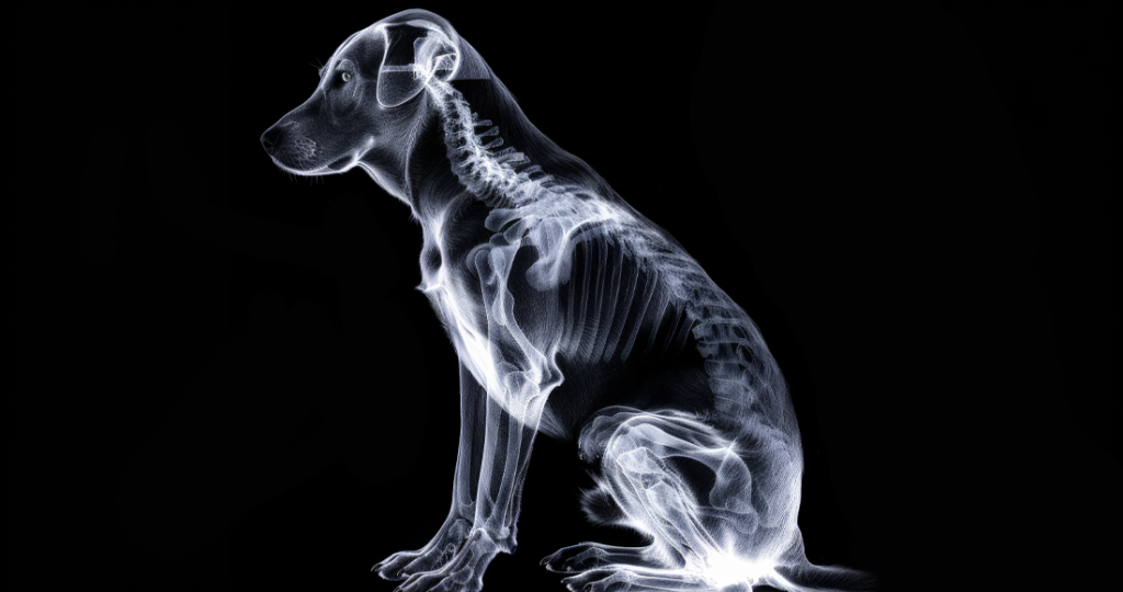
Dental radiography has revolutionized veterinary dentistry by revealing the two-thirds of tooth structure hidden beneath the gum line where most dental disease originates and progresses unseen. Professional pet dental x-rays Fairfield TX services at our veterinary clinic utilize state-of-the-art digital radiography equipment that provides immediate, high-resolution images essential for accurate diagnosis and treatment planning. Without these crucial images, veterinarians are essentially working blind, missing up to 40% of dental pathology that exists below the visible crown.
The importance of dental radiographs cannot be overstated, as numerous studies demonstrate that visual examination alone fails to identify significant disease requiring treatment. Teeth that appear healthy externally may harbor abscesses, root fractures, or resorptive lesions causing significant pain. Conversely, teeth that look severely diseased might have surprisingly healthy roots worth preserving with appropriate treatment.
Modern digital dental radiography offers numerous advantages over traditional film, including immediate image availability, superior image quality, reduced radiation exposure, and the ability to enhance images for better visualization. This technology has made comprehensive dental radiography the standard of care, transforming it from an optional diagnostic tool to an essential component of every professional dental procedure.
Understanding Dental Radiograph Necessity
Every professional dental cleaning should include full-mouth radiographs to establish baseline images and identify hidden pathology requiring treatment. The American Veterinary Dental College considers dental radiography mandatory for proper diagnosis and treatment planning. Attempting dental procedures without radiographs is comparable to performing surgery without being able to see the surgical field.
Clinical signs often fail to correlate with radiographic findings, as pets instinctively hide pain and continue eating despite significant oral disease. A cat with severe tooth resorption may show no outward signs while experiencing constant discomfort. Dogs with root abscesses might only exhibit subtle behavioral changes that owners attribute to aging rather than dental pain.
The investment in dental radiography pays dividends through accurate diagnosis leading to appropriate treatment, preventing unnecessary extractions of salvageable teeth while ensuring diseased teeth are removed. This targeted approach reduces anesthesia time, surgical trauma, and ultimately provides better outcomes. Our pet dental x-rays Fairfield TX capabilities ensure no pathology goes undetected.
Digital Radiography Technology and Advantages
Digital dental radiography uses electronic sensors instead of traditional film to capture images, similar to how digital cameras replaced film photography. When x-rays pass through teeth and surrounding structures, the digital sensor converts the radiation into electronic signals that create detailed images on a computer screen within seconds. This immediate feedback allows repositioning if needed without waiting for film development.
The superior image quality of digital radiographs reveals subtle changes in tooth and bone density that might be missed on traditional film. Software enhancement tools allow adjustment of contrast, brightness, and magnification without retaking images. Measurement tools provide precise evaluation of periodontal bone loss, root canal width, and other critical parameters.
Radiation exposure with digital systems is 60-90% less than traditional film radiography, improving safety for both patients and staff. The increased sensitivity of digital sensors requires less radiation to produce diagnostic quality images:
- Immediate image availability for evaluation
- No chemical processing eliminating environmental concerns
- Easy image storage and retrieval for comparison
- Simple sharing with specialists for consultation
- Enhanced visualization through digital manipulation
- Reduced radiation exposure for patients
- Efficient workflow reducing anesthesia time
Comprehensive Dental Radiograph Series
Full-mouth dental radiographs require multiple images to visualize all teeth and surrounding structures adequately. Dogs typically need 10-14 radiographs while cats require 6-10, depending on skull conformation and missing teeth. Each image captures specific teeth using precise angles that minimize distortion and overlap.
Intraoral radiography, where the sensor is placed inside the mouth, provides the highest quality images with minimal distortion. Parallel technique works well for mandibular teeth where the sensor can be positioned parallel to the tooth roots. Bisecting angle technique is necessary for maxillary teeth where palate anatomy prevents parallel placement.
Extraoral techniques may be necessary for caudal teeth or when intraoral placement isn’t possible due to anatomy or pathology. While these views provide valuable information, they typically have more distortion and overlap than intraoral images. Combining techniques ensures complete visualization of all dental structures.
Common Pathology Revealed by Dental X-Rays
Periodontal disease progression becomes clearly visible on radiographs through horizontal and vertical bone loss patterns around tooth roots. Early bone loss appears as subtle changes in bone density, while advanced disease shows dramatic bone destruction. Serial radiographs document disease progression or improvement with treatment, guiding ongoing management decisions.
Root abscesses appear as dark halos around root tips where infection has destroyed surrounding bone. These painful lesions often develop from tooth fractures, severe periodontal disease, or endodontic disease. Without radiographs, these infections continue spreading, potentially creating facial swellings or draining tracts.
Tooth resorption, particularly common in cats, shows as moth-eaten appearances where tooth structure is being destroyed by the body’s own cells. These intensely painful lesions often hide below the gum line, invisible without radiographs. Early detection allows extraction before severe pain develops, significantly improving patient comfort.
Fractured Teeth and Root Evaluation
Dental radiographs reveal the extent of tooth fractures and help determine treatment options between extraction and root canal therapy. Crown fractures may extend below the gum line into root structure, information crucial for treatment planning. Root fractures might be present without visible crown damage, causing pain with normal appearance.
Pulp exposure assessment through radiographs evaluates whether the sensitive inner tooth remains vital or has become infected. Changes in pulp canal width compared to adjacent teeth indicate tooth death requiring treatment. Periapical changes suggest infection spreading beyond the tooth into surrounding bone.
Previous dental work evaluation benefits from radiographs showing root canal fills, post placement, or retained root tips from previous extractions. These findings influence current treatment decisions and explain ongoing problems. Our pet dental x-rays Fairfield TX services include thorough evaluation of all previous dental work.
Developmental and Congenital Abnormalities
Missing teeth require radiographic confirmation to differentiate between congenitally absent teeth and embedded or impacted teeth requiring treatment. Embedded teeth can form dentigerous cysts that destroy surrounding bone if not removed. What appears as a missing tooth might hide significant pathology below the surface.
Supernumerary (extra) teeth create crowding leading to increased periodontal disease and malocclusion. Radiographs reveal whether extra teeth have normal root structure or are fused with adjacent teeth. This information guides extraction decisions and techniques.
Developmental abnormalities including enamel hypoplasia, abnormal root formation, or tooth fusion become apparent on radiographs. These conditions predispose teeth to disease and may require preventive extraction or special monitoring. Early identification allows appropriate management before complications develop.
Pre-Extraction Planning and Assessment
Radiographs before tooth extraction reveal root number, shape, and orientation crucial for planning surgical approach. Curved, divergent, or dilacerated roots require sectioning and careful elevation to prevent fracture. Ankylosed teeth fused to bone need extensive bone removal for extraction.
Adjacent structure evaluation prevents iatrogenic damage during extraction by revealing proximity to mandibular canal, nasal cavity, or adjacent tooth roots. This information guides surgical planning to avoid complications. Knowledge of anatomical relationships improves surgical outcomes and reduces complications.
Post-extraction radiographs confirm complete tooth removal, as retained root tips can cause ongoing infection and pain. Even experienced surgeons occasionally leave small root fragments that only radiographs reveal. Immediate recognition allows prompt removal before healing begins.
Oral Mass and Tumor Assessment
Radiographic evaluation of oral masses reveals bone involvement that influences prognosis and surgical planning. Benign masses typically show smooth, well-defined borders with minimal bone involvement. Malignant tumors often display irregular borders with extensive bone destruction.
Biopsy planning benefits from radiographic guidance showing the extent of bone involvement and helping select representative sampling sites. This information influences whether incisional or excisional biopsy is appropriate. Understanding tumor extent before surgery improves margin planning.
Treatment planning for oral tumors requires detailed radiographic assessment to determine surgical margins needed for complete excision. Some tumors appearing small externally show extensive bone invasion requiring aggressive surgery. Our pet dental x-rays Fairfield TX capabilities ensure accurate tumor staging.
Jaw Fracture and Trauma Evaluation
Mandibular and maxillary fractures require radiographic evaluation to determine fracture configuration, displacement, and involvement of tooth roots. This information guides treatment decisions between conservative management and surgical repair. Multiple views may be necessary to fully evaluate complex fractures.
Temporomandibular joint evaluation through radiographs reveals luxations, fractures, or degenerative changes affecting mouth opening and comfort. These problems cause difficulty eating and chronic pain if not properly diagnosed and treated. Comparison views of both joints identify subtle asymmetries.
Previous trauma assessment benefits from radiographs revealing old fractures, retained foreign bodies, or chronic osteomyelitis. These findings explain ongoing problems and guide current treatment. Historical changes influence prognosis and treatment options.
Feline-Specific Radiographic Findings
Tooth resorption affects up to 70% of cats over five years old, with radiographs essential for diagnosis and treatment planning. Different types of resorption require different treatment approaches, information only available through radiography. Complete examination reveals multiple affected teeth requiring treatment.
Feline chronic gingivostomatitis evaluation includes radiographs to assess bone involvement and identify retained root fragments contributing to inflammation. Full-mouth extractions often necessary for this condition require radiographic guidance. Post-extraction radiographs ensure complete removal of all root fragments.
End-stage kidney disease in cats can cause significant jaw bone changes visible on dental radiographs, including rubber jaw from calcium depletion. These findings influence anesthetic protocols and extraction techniques. Recognition prevents iatrogenic jaw fractures during procedures.
Monitoring and Follow-Up Radiography
Serial radiographs document treatment response and disease progression, particularly valuable for periodontal disease management. Comparing current images to previous radiographs reveals subtle changes not apparent on single examinations. This monitoring guides treatment modifications and helps determine extraction timing.
Endodontic treatment follow-up requires radiographs to assess healing and identify complications like root canal failure or persistent infection. Standard protocols include immediate post-treatment radiographs and follow-up imaging at 6-12 months. Long-term monitoring ensures treatment success.
Orthodontic treatment monitoring through sequential radiographs ensures teeth move appropriately without causing root damage. These images guide appliance adjustments and determine treatment endpoints. Documentation provides evidence of successful treatment completion.
Integration with Treatment Planning
Radiographic findings directly influence treatment decisions, often dramatically changing planned procedures based on visual examination alone. Teeth appearing suitable for cleaning might require extraction based on radiographic findings. Conversely, teeth looking hopeless might be salvageable with appropriate treatment.
Cost estimates become more accurate with radiographic information allowing precise treatment planning. Owners can make informed decisions about which teeth to treat, extract, or monitor. This transparency builds trust and ensures optimal resource allocation.
Treatment prioritization based on radiographic severity ensures the most painful conditions receive immediate attention. Limited budgets can focus on teeth causing greatest discomfort. Our pet dental x-rays Fairfield TX services provide information necessary for these important decisions.
Safety Protocols and Radiation Protection
Modern digital dental radiography significantly reduces radiation exposure, but proper safety protocols remain essential. Lead aprons and thyroid shields protect patients from scatter radiation. Proper collimation limits radiation to necessary areas only.
Staff safety involves maintaining distance during exposures, using protective equipment, and following proper positioning techniques. Dosimetry badges monitor cumulative exposure ensuring safe levels. Regular equipment maintenance ensures optimal performance with minimal radiation.
Pregnant staff members avoid radiography areas during exposures as standard precaution despite minimal risk with proper protocols. Alternative duties during pregnancy eliminate any potential concerns. Safety remains paramount in all radiographic procedures.
Quality Assurance and Image Optimization
Proper positioning and technique are crucial for diagnostic quality images that accurately represent dental pathology. Regular training ensures consistent high-quality radiographs. Standardized protocols minimize retakes and reduce radiation exposure.
Image processing optimization involves adjusting contrast, brightness, and filters to enhance visualization of specific structures. Different settings work best for evaluating teeth versus bone. Experience interpreting dental radiographs improves diagnostic accuracy.
Equipment maintenance including regular calibration ensures consistent image quality and proper radiation output. Sensor care prevents artifacts that could obscure pathology. Investment in quality equipment and maintenance provides reliable diagnostic imaging.
Schedule Your Pet’s Dental Radiographs
Don’t let hidden dental disease cause your pet unnecessary pain and suffering. Contact our Fairfield veterinary clinic at 1501 W US Highway 84 to schedule comprehensive dental evaluation including digital radiographs. Our pet dental x-rays Fairfield TX services reveal problems invisible during routine examination.
Every professional dental cleaning at our clinic includes full-mouth radiographs ensuring complete diagnosis and appropriate treatment. We’ll show you the images and explain findings so you understand your pet’s oral health status. This transparency helps you make informed decisions about treatment options.
Trust our advanced digital radiography to provide the detailed images necessary for optimal dental care. Your pet deserves comprehensive diagnosis revealing all dental pathology, not just what’s visible on the surface. Schedule their dental radiographs today and ensure their mouth is truly healthy.